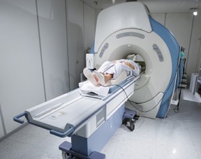
A computed tomography (CT) scan (also known as a computed axial tomography scan, or CAT scan) is one of the most commonly used tools for the screening, diagnosis and treatment of prostate cancer. A CT scan uses X-rays and computers to produce three-dimensional, cross-sectional images of inside the human body. However unlike traditional X-rays, CT scans provide remarkably detailed images of the bones, organs and tissues. CT scans can also show a tumor’s shape, size, and location.
Preparation Before The Scan
Some CT scans may require the use of contrast dye which can be administered either intravenously, swallowed or put into the intestines through the rectum as an enema prior to the procedure. The contrast dye is used to create clearer images of the prostate . Before receiving the dye however, be sure to let your healthcare team know if you’ve ever had an adverse reaction to contrast dye, seafood, or iodine. If there’s a risk of having an allergic reaction, you may be given a test dose of the contrast dye first before receiving additional contrast.
How It Works
A CT scan uses a pencil-thin beam to create a series of images taken from varying angles.The information from each angle is then fed into a computer, which then creates a black and white picture that shows a slice of a certain area of the body. The cross-sectional images are like a slice of bread taken from a loaf. By layering the CT image slices on top of one another, the machine can create a 3-dimensional (3-D) view of the prostate. The 3-D image can be rotated on a computer screen to look at it from different angles.
A CT scan is not only used to pinpoint the location of a tumor but can also be used to evaluate the extent of cancer in the body, and assess whether the disease is responding to treatment. In some instances, CT technology can also be used to accurately guide cancer treatment.
A CT scan can also reveal blood flow and the anatomy of tissues in and around the prostate, allowing for the diagnosis and monitoring of tumor growth.
Potential Side Effects
Side effects from the contrast dye can vary among patients and can include:
- Rash
- Nausea
- Wheezing
- Shortness of breath
- Itching or facial swelling that can last up to an hour
Side effects of contrast dye are usually mild and will often go away on their own. However a side effect can be a sign of a more serious reaction that needs to be addressed. Be sure to let your radiology technologist and your health care team know if you notice any physical changes after receiving the contrast dye.


